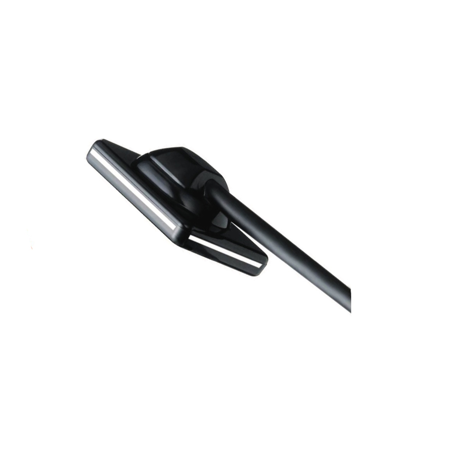
Televere Systems SOPIX Veterinary Dental Sensors
SOPIX sensors surpass the limits of radiological examinations by offering greater differentiation of dental tissue. This technological achievement is called FIBER2PIXEL. Striking contrast for a more reliable diagnosis. Differentiation of the dental tissue FIBER2PIXEL® technology is based on the use of broad spectrum optical microfibers for the guided transmission of photon emissions in order to provide highly contrasted images.
Related products
-
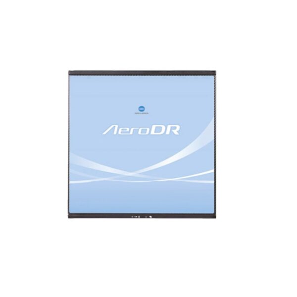
Konica Minolta AeroDR
Avoid unplanned downtime and achieve greater productivity with the reliable AeroDR XE. Our lithium ion capacitor extends the panel’s life to last an entire shift up to 8.2 hours or 300 images. New panel drop sensors and monitoring provides ongoing data on panel handling, so you can avoid catastrophic failures, reduce repair costs and gain peace of mind. With the Aero Remote, you can even perform initial diagnostic checks without calling for support. Plus, as the lightest panel on the market at 5.7 lbs, the AeroDR XE is easier to handle with convenient grip strips.
-
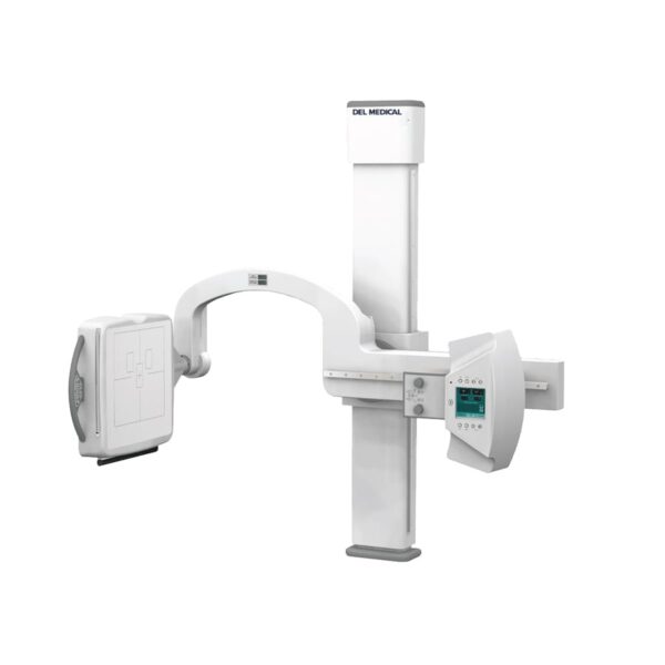
UMG/Del Medical U-Arm System
Del Medical UArm Systems are designed for use in hospital emergency rooms, orthopaedic and all general radiographic applications. The UArm maintains constant alignment between the x-ray tube and image receptor, regardless of tilt position or image receptor angle. The extraordinary flexibility of this system makes it ideal for all radiographic procedures including sitting, erect and recumbent positions.
-
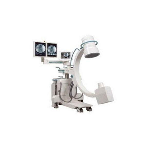
Ziehm Quantum
– Single Function Foot Switch
– Real Time Processing
– Filters/Windowing/Rotation/Mirroring
– Post Processing-Edge Enhancement
– Rotation, Windowing, Inverse, Image Crop
– High Frequency X-ray Generator 20,000 Hz
– Iris and Slot During live Fluoroscopy
– Fluoroscopic Operation from 40kV to 110kV at 0.2-Maximum 6ma and Pulse Modes
– Radiographic Operation from 40kV to 110kV at Maximum 20ma
– 8ma Mode for Digital Radiograph/Snap Shot Mode
– Dual Mode 9/6 in. Image Intensifier
– High Contrast Camera
– Body Region and Application Specific Key, Extremities, Chest, head, Spine, Hip Metal, Soft Tissue, 1/2 Dose
– Large Patient Diameter key
– Integrated Cable Pusher Allow for Easy Maneuverability and Positioning
– Two 18 in. High Resolution/Brightness TFT Flat Panel Monitors, 1280×1024
– 1280x1024Vx12 bit Highline Video Image Display with Dual Graphic Overlays
– 360 Degree Digital Continuous Image Rotation
– Last Image Hold
– Invert, White on Black
– True 1024 Shades of Gray
– Real Time Noise Reduction (Low, Medium, High)
– Snapshot, Electronic Noise Reduction by Multiple Frame Integration Preset for Better Image Quality
– Touch Screen Text Keyboard for Patient Annotation
– Auto Store Feature
– True 16 bit Image Processing and Storage
– 10,000 Digital Image Stored in 1Kx16 bit Image Display -
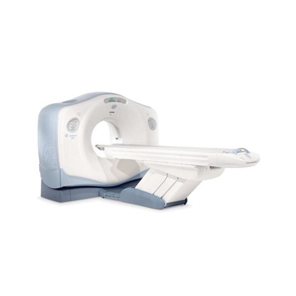
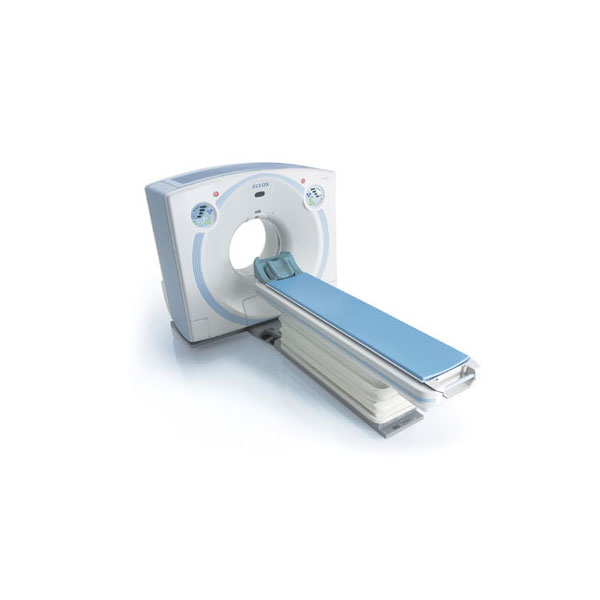
Hitachi Airis
For both you and Hitachi, economy, patient comfort and confidence in diagnosis are vital. AIRIS Vento incorporates newly developed permanent open MRI technologies and a wealth of the latest know-how in superconductive MRI systems to provide you with enhanced imaging capabilities and an outstanding return on investment.
-
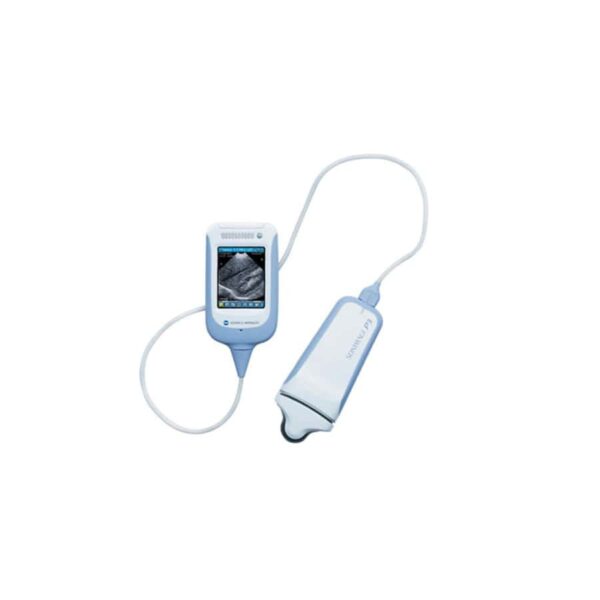
Konica Minolta SONIMAGE P3
The SONIMAGE P3 is a true portable ultrasound machine that gives clinicians the ability to do more for patients where and when they need it most – at the point-of-care. With its small footprint and weighing less than a pound, this handheld device can accelerate and improve interventions and decision making time.
Intuitively designed like a smart-phone, the P3 equips users with a non-invasive tool to see what’s beneath the surface in patients, giving care providers’ quicker access to information and improving patient care. The SONIMAGE P3, cost-effectively empowering physicians to do more with less – providing them imaging solutions in real time.
-
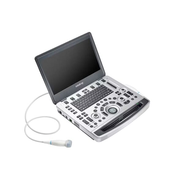
MindRay M9
Based on Mindray’s new generation ultrasound platform, mQuadro, M9 has raised the industry standards to an all new level. Advanced signal transmission and reception processors provide highly sensitive and accurate echo detection. Innovative transducer technologies allow for better penetration, higher resolution, greatly enhancing your diagnostic experience.

